Cone beam imaging is transforming the way dentists collate information about your oral health. It’s also changing the way that our dentists here at AZ Family Dental gather information and carry out dental procedures to improve your smile.
In the future you might be scheduled for a dental cone beam CT scan if you require a dental X-ray. Utilizing a cone beam CT scan provides you with a more improved clinical experience, in addition to just using traditional x-raying methods. It’s a superior tool used to rapidly diagnose and work out a treatment plan regardless of your dental needs.
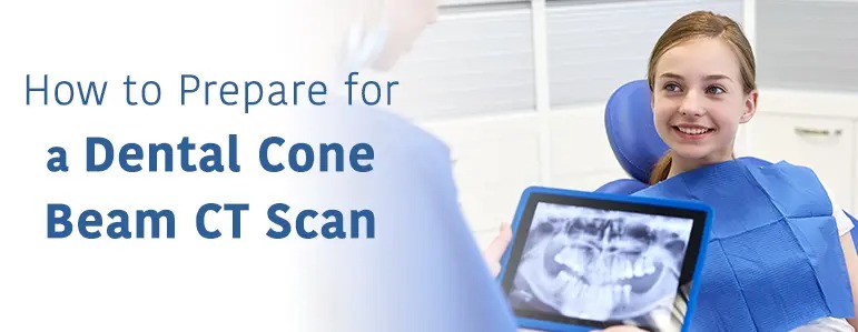
As this technology is relatively new, it’s natural to be curious about what a dental cone beam CT scan might entail. Don’t worry, these scans are painless and only take seconds. In this guide, we help you to learn more about what a dental cone beam CT scan is, the benefits of this cutting-edge 3D technology, and what you can expect.
One of the dentists here at AZ Family Dental may have advised you that you need to have a cone beam scan. This is exciting, as a dental cone beam CT scan, otherwise known as Cone Beam Computed Tomography (CBCT), is a diagnostic scan that utilizes 3D dental imaging.
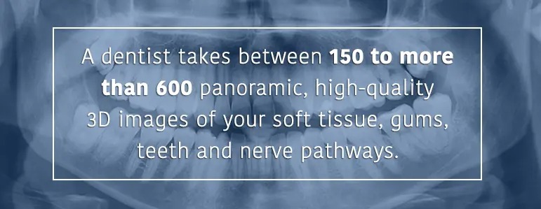
3D imaging is an impressive development in dental diagnostic technology and gives superior views over antiquated 2D imaging technology. Using a computer, X-rays, and a single scan, one of our dentists takes between 150 to more than 600 panoramic, high-quality 3D images of your soft tissue, gums, teeth and nerve pathways.
Dental cone beam CT scans provide clear 3D images of the complexity of the maxillofacial region. The scan collates images for a complete dental evaluation or review of a small section of your facial bones. A CBCT machine, like the Sirona Orthophos SL-AI used here at AZ Family Dental, involves approximately 1,000 X-rays compiled together to render a 3D skull.
Conventional medical computerized tomography scans, better known as CT, take images in slices or via a continuous spiral motion. CBCT is different in that it sweeps past the target area once or twice. It shares a similarity to panoramic radiography in this sense.
The X-ray energy involved in CBCT is quite similar to that found in conventional medical computerized tomography too. The operational differences is what makes them stand apart. Sometimes people get the two confused as both provide 3D images and include the term computed tomography.
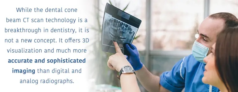
While the dental cone beam CT scan technology is a breakthrough in dentistry, it is not a new concept. It offers 3D visualization and much more accurate and sophisticated imaging than digital and analog radiographs. Although it was first devised as an efficient and cost-effective way of obtaining 3D cross-sectional images for radiotherapy, it was also later used in angiography.
Until the introduction of 3D imaging, the concepts of dental radiography didn’t change significantly from the earliest days of dental radiographs. Computed tomography was available for 3D imaging in the ‘80s In 1988, cone beam computerized tomography begun to be used in dentistry, according to the report The Genesis and Development of CBCT for Dentistry. However, due to cost, the technology was only used in unique dental situations, such as complex surgeries and craniofacial anomalies.
A cone beam 3D imaging scan provides the dentists with particularly detailed information that can’t be obtained from regular X-rays. For instance, it enables dental professionals to assess the exact shape and thickness of your bones if you’re being considered for certain dental procedures, such as dental implants.
The impact that CBCT technology has had on dental imaging shouldn’t be underestimated. Dental cone beam 3D imaging enables the dentists to give a rapid and accurate diagnosis. They can then better prepare for your dental procedure.
Common uses for the dental cone beam CT scan include:
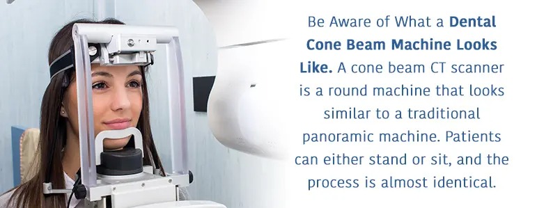
A cone beam CT scanner is a round machine that looks similar to a traditional panoramic machine. Patients can either stand or sit, and the process is almost identical.
The arm rotates 360degrees around your head, taking only a few seconds. While doing so, it captures multiple images through a cone beam from a variety of angles. These are then finally put together to create one single 3D image for the dentists to review.
CBCT scans give us a full view of the 3D structure, however, they are not used for diagnosing cavities. Traditional dental X-rays are still needed for cavity detection and are considered standard of care. Both methods are equally important, serve different purposes, and are used in modern dentistry.
There may not be other alternatives if the dentists have decided that you require a CBCT scan. Traditional dental X-rays may not allow the dentists to assess your bone accurately enough to treat you safely.
It’s important to note the differences between the CBCT and CEREC (Chairside Economical Restoration of Esthetic Ceramics) technology.
The CBCT scan creates a 3D image used for treatment planning and diagnosing. The CBCT is used to better diagnose pathology, infections, bone conditions implant planning, etc.
On the other hand, the CEREC machine is the technology that takes the digital impression. It is used to fabricate the actual restorations (inlays, onlays and crowns).
CEREC and CBCT 3D technology can be combined to more accurately plan and fabricate dental improvements. They each have their roles individual and are not the same thing. Dental restorations or appliances are not made with the CBCT alone.
The beauty of the CEREC and CBCT machines is the seamless integration between the two to provide better dental care to our patients.
Conventional X-rays are flat, 2D pictures, that don’t provide the dentists with the best information for dental diagnostics and planning.
Dental CBCT scans are non-invasive, and a fast method of diagnosis. Other benefits include:
CT scanning is accurate and painless.
Focused X-ray beam results in improved image quality as it reduces scatter radiation.
You shouldn’t anguish about what to expect from dental CBCT scan. You also don’t need to worry about being in an enclosed space like some other types of CT scans.
Depending on the exact type of scanner being used, you will either be asked to stand or sit in the exam chair. The dentist positions you so the area they wish to look at is centered in the beam.
The machine rotates around your face and head. You may have already seen panoramic dental radiography units in dental practices and hospitals, and the CBCT scan machine moves similarly.
While the detector and X-ray source revolves around you, you’ll need to be very still. You’ll perhaps be asked to grasp the machine’s handles, so you stay in the same place and remain comfortable. With your dental CBCT scan, you don’t need to bite down on an uncomfortable piece of hard plastic, which is beneficial for many clients, especially pediatric patients.
For a full mouth X-ray, the actual CBCT scan examination takes only between 20 to 40 seconds to complete. A regional scan focusing on one particular area takes less than 10 seconds on average.
You must tell the dentists if there’s the possibility you’re pregnant. How to prepare for a CBCT X-ray starts by visiting the dental office at your appointed time. You don’t need to do anything special or different to your usual routine beforehand. You will usually need to wear a gown.
There are several items that you need to remove before your exam, including
You may need to wear a piece of equipment called a scanning appliance during your scan if you’re having a CBCT scan to plan your dental implants. This is a special plate you wear much the same way as a denture. It contains markers that guide your examination and only needs to be worn during your scan.
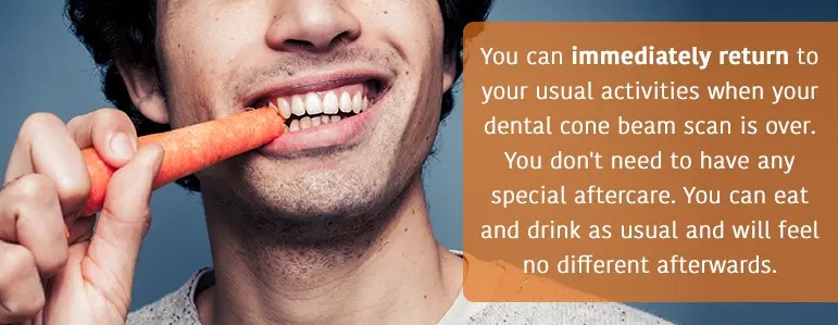
You can immediately return to your usual activities when your dental cone beam scan is over. You don’t need to have any special aftercare. You can eat and drink as usual and will feel no different afterwards.
Your dentist will then analyze your results. You’ll then find out what treatment plan or method is best for you.
To recap, the benefits of dental cone beam CT scans are:
This advanced technology will shape the future of dental diagnosis and treatment planning. The advantages of 3D imaging apply to all dental disciplines. It, therefore, may be integrated it into more dental offices in the future.
According to the report, What is Cone-Beam CT and How Does it Work,it’s thought that dental cone beam scan use will increase over the coming years although CBCT dentistry is a relatively new field. Experts believe that in the future, scan times will be reduced even further.
Keep in mind, there are some things digital X-rays are better for, and there are other things CBCT images are better for. Both are needed for state-of-the-art dentistry.
A dental cone beam CT scan is easy to prepare for and is a quick and painless way for the dentists here at AZ Family Dental to assess your mouth health and plan reconstructive and cosmetic dentistry improvements.
AZ Family Dental is committed to providing our patients with advanced technology and medical advances, including 3D technology. If you have any questions about the Dental Cone Beam CT Scan or other equipment and technology we utilize here at AZ Family Dental, call our team at 623-777-2037.